Bovine Collagen Type Composition: Type I, Type II, and Type III Explained
In this article, we will explore the significance of bovine collagen type 1 2 3 in various applications.
Standard bovine collagen peptides derived from hide and bone contain Type I and Type III collagen — not Type II. This is the single most important fact about bovine collagen type composition. If the product comes from conventional bovine sources such as skin, bone, or tendon, the collagen is predominantly Type I with a smaller proportion of Type III. Type II collagen is found exclusively in cartilage and is absent from hide- or bone-derived collagen peptides. 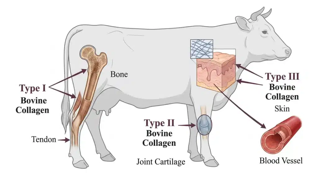
Understanding bovine collagen type composition means understanding three separate proteins with distinct molecular structures, tissue origins, and biological roles. This guide breaks down each type — what it is, where it comes from, and how the proportions vary across different bovine tissues.
The Molecular Foundation of Bovine Collagen Types
Collagen is the most abundant protein in mammals, comprising 25 to 35 percent of total body protein. The collagen molecule consists of three polypeptide chains coiled around one another in a triple helix. Bovine collagen types are classified according to the specific chain composition of this helix.
The chain composition determines not only the molecular structure but also the tissue distribution and biological function of each collagen type. Three types are relevant to bovine sourcing:
| Collagen Type | Chain Composition | Primary Bovine Tissue Sources | Biological Role |
|---|---|---|---|
| Type I | Heterotrimer: two α1(I) chains + one α2(I) chain | Bone, tendon, skin, ligament, blood vessel walls, cornea | Provides tensile strength; structural framework for bone and connective tissue |
| Type II | Homotrimer: three α1(II) chains | Articular cartilage, intervertebral discs, vitreous humor | Enables cartilage to withstand compression and absorb shock |
| Type III | Homotrimer: three α1(III) chains | Skin, blood vessels, internal organs, uterus, periodontal ligament | Contributes to tissue elasticity and vascular integrity |
Type I is a heterotrimer — its three chains are not identical. Type II and Type III are homotrimers — each consists of three identical chains. This molecular distinction underpins everything else about how these collagens behave in the body.
Type I Collagen: The Predominant Bovine Collagen
Type I collagen constitutes approximately 90 percent of total body collagen in cattle and other mammals. It is the primary structural protein in bone, tendon, skin, and ligaments — tissues that must resist stretching and mechanical load.
Molecular structure. The heterotrimeric chain composition of Type I — two α1(I) chains and one α2(I) chain — produces thick, densely packed fibers with exceptional tensile strength. The amino acid sequence is rich in glycine, proline, and hydroxyproline, which stabilize the triple helix and contribute to thermal stability.
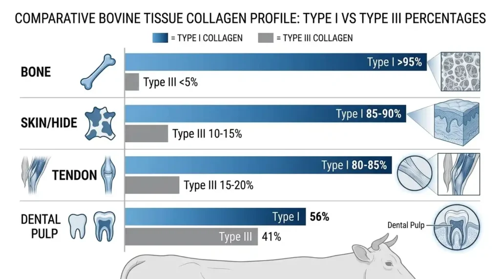
Tissue distribution. In bovine bone, Type I collagen accounts for over 95 percent of the collagen content. In bovine skin, the proportion is approximately 85 to 90 percent. In bovine knee meniscus fibrocartilage, the pepsin-solubilized collagen fraction is approximately 98 percent Type I. In bovine synovial capsule, Type I makes up 83 percent of total collagen.
Functional significance. Type I collagen provides the organic matrix upon which bone mineralization occurs. In tendons and ligaments, it transmits mechanical force from muscle to bone. In skin, it forms the dermal scaffold that maintains structural integrity. For products targeting bone health, skin structure, or wound healing, Type I is the active component.
Type II Collagen: The Cartilage-Specific Type — Why It Is Not Present in Standard Bovine Peptides
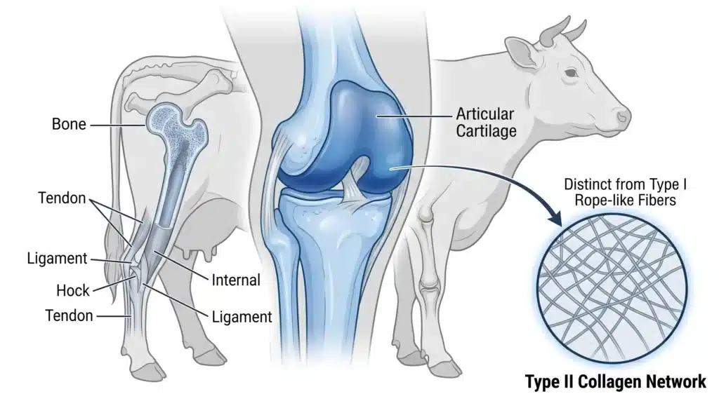
Type II collagen has a fundamentally different tissue distribution than Type I and Type III. It is restricted to cartilage and related tissues, which means it does not appear in the hides, bones, or tendons that serve as raw materials for standard bovine collagen peptides.
Molecular structure. Type II collagen is a homotrimer of three α1(II) chains. The fibers it forms are thinner than Type I fibers and organize into a loose, mesh-like network. This architecture allows cartilage to deform under compression and return to its original shape — an essential property for load-bearing joints.
Tissue distribution. Type II collagen is the dominant collagen in bovine articular cartilage, intervertebral discs, and nasal-septum cartilage. In bovine vitreous humor, Type II is also a major component. In the inner medial meniscus of cattle, Type II accounts for approximately 60 percent of the collagen, with Type I making up the remaining 40 percent. In bovine retinal microvessels, Type II collagen has been identified as a component of the extracellular matrix.
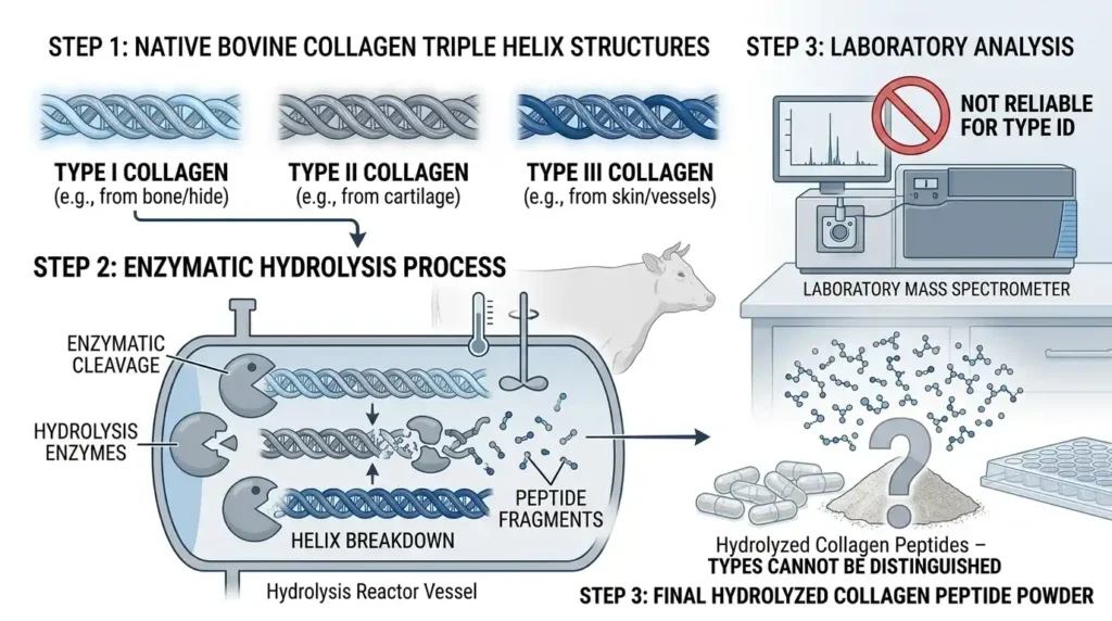
Why standard bovine collagen peptides do not contain Type II. The hides and bones used for conventional bovine collagen peptide production are rich in Type I and Type III — they contain no Type II. To obtain Type II collagen, the raw material must be cartilage — typically bovine tracheal cartilage or chicken sternum — extracted through a different process. If a product is labeled simply as “bovine collagen peptide” without specifying cartilage as the source, it does not contain Type II.
Type III Collagen: The Companion to Type I
Type III collagen frequently co-localizes with Type I in the same tissues. This is why standard bovine collagen peptides naturally contain both — the proportion depends on the specific tissue source.
Molecular structure. Type III collagen is a homotrimer of three α1(III) chains. It forms thinner, more flexible fibers than Type I. This flexibility makes Type III particularly important in tissues that require both structural support and elasticity.
Tissue distribution. In bovine skin, Type III accounts for approximately 10 to 15 percent of the collagen — the remainder is Type I. In bovine periodontal ligament, about 20 percent is Type III. In bovine dental pulp, Type III represents 41 percent of the collagen, while Type I accounts for 56 percent and Type V accounts for 2 percent. In bovine synovial capsule, Type III makes up 17 percent of the collagen not accounted for by Type I.
Functional significance. Type III collagen supports the elasticity of blood vessels and internal organs. It is also present in the skin, where it works alongside Type I to maintain both strength and suppleness. Because Type III is naturally present in bovine hide, hide-derived collagen peptides always include it as a minor but meaningful component.
Bovine Collagen Type Proportions: How Percentages Vary by Tissue
The proportion of Type I to Type III collagen is not fixed — it varies significantly depending on the tissue source. The following table summarizes the collagen type composition of various bovine tissues:
| Bovine Tissue | Type I Collagen | Type III Collagen | Other Collagen Types |
|---|---|---|---|
| Bone | >95% | <5% | Trace |
| Skin (hide) | 85–90% | 10–15% | Trace |
| Tendon | 80–85% | 15–20% | Trace |
| Ligament | 80–85% | 15–20% | Trace |
| Periodontal ligament | ~80% | ~20% | — |
| Dental pulp | 56% | 41% | 2% Type V |
| Synovial capsule | 83% | 17% | — |
| Meniscus (outer region) | >80% | <10% | Trace Types III, V |
Key observations from these data:
- Bone-derived collagen peptides are almost entirely Type I, with minimal Type III.
- Skin-derived collagen peptides consistently contain both Type I and Type III, with Type I in the majority.
- Some tissues, such as dental pulp, are significantly richer in Type III — but these are not typical sources for commercial collagen peptide production.
- The Type I/III ratio is a direct reflection of the tissue source, not a quality indicator.
Commercial bovine hide collagen preparations are typically approximately 97 percent Type I with the remainder comprised of Type III collagen. This ratio is consistent across most commercial products because hide is the dominant raw material.
Structural and Functional Comparisons Among Bovine Collagen Types
Beyond tissue distribution and proportion, the three collagen types differ in physical and biochemical properties:
Fiber morphology. Type I fibers are thick, densely packed, and highly resistant to tensile forces. Type II fibers are thinner and form a loose network suited to compressive loading. Type III fibers are thin and flexible, often interwoven with Type I fibers to provide a balance of strength and elasticity.
Charge characteristics. Titration studies of collagen Types I, II, and III between pH 7.0 and 2.0 have shown that Type I collagen has the most titratable carboxylic groups. Type II and Type III have significantly fewer titratable groups. This difference influences how each collagen type interacts with glycosaminoglycans and other extracellular matrix components.
Platelet aggregation activity. Bovine Type I, Type II, and Type III collagen differ in their ability to induce human platelet aggregation. Type III collagen is a particularly potent inducer, while Type II shows weaker activity. This distinction is relevant for applications in wound healing and hemostasis.
Thermal stability. Type I collagen generally has a slightly higher thermal denaturation temperature than Type III. Type II, with its higher hydroxylysine content, exhibits distinct stability properties. However, once collagen is hydrolyzed into peptides — as it is in commercial products — these thermal differences are largely irrelevant because the triple helix has been disrupted.
The Mislabeling Issue: Claims of “Type I, II, and III” in Bovine Products
A widespread problem in the collagen industry is the labeling of bovine products as containing “Types I, II, and III.” This claim is often inaccurate.
What the claim implies. A product labeled as containing bovine Type I, II, and III collagen suggests that it provides all three types from bovine sources. For this to be true, the product must contain both standard hide- or bone-derived collagen (Type I and III) and cartilage-derived collagen (Type II).
What is actually in the product. Most products making this claim do not contain cartilage-derived collagen at all. They contain only hide- or bone-derived collagen — which is Type I and III — and the Type II claim is either speculative or false.
Why verification is difficult. Once collagen is hydrolyzed into peptides, the distinguishing molecular features of each collagen type are largely lost at the macroscopic level. Conventional analytical methods such as amino acid analysis, SDS-PAGE, and infrared spectroscopy cannot reliably differentiate Type I from Type II or Type III in hydrolyzed samples. Verification requires mass spectrometry or enzyme-linked immunosorbent assays — methods that are expensive and rarely performed in routine quality control.
What to look for. When specifying bovine collagen peptides, the key question is not whether the product contains Type I, II, and III — it is what tissue source was used. Hide- and bone-derived products contain only Type I and III. Cartilage-derived products contain Type II. A product that claims to contain all three must document both the tissue sources and the blending ratios.
Application Relevance of Bovine Collagen Type Composition
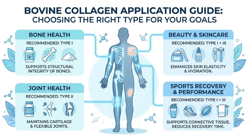
The collagen type composition of a product directly influences its suitability for different applications:
| Application | Relevant Collagen Types | Rationale |
|---|---|---|
| Bone health supplements | Type I (bone source) | High Type I content supports bone matrix and mineralization |
| Skin health/beauty supplements | Type I + III (hide source) | Both types are present in skin; Type I provides structure, Type III provides elasticity. |
| Joint health supplements | Type II (cartilage source) | Type II is the structural protein of articular cartilage |
| Tendon and ligament support | Type I + III | Both types are present in these connective tissues |
| Wound care / hemostatic products | Type I + III (specific ratios) | Type III is more active in platelet aggregation |
| Functional beverages | Type I + III (low molecular weight) | Good solubility and neutral flavor make hide-derived peptides suitable |
The choice of collagen type should be driven by the intended biological target. A product formulated for joint health should include Type II from cartilage. A product formulated for skin should include Type I and III from hide. Using a hide-derived product for joint health misses the target — and using a cartilage-derived product for skin health is similarly misaligned.
Final Summary: What Matters About Bovine Collagen Type Composition
Three points define the practical reality of bovine collagen type composition:
- Standard bovine collagen peptides contain Type I and Type III, not Type II. This is a function of the raw material — hide and bone contain no Type II. Type II is found only in cartilage and requires separate sourcing.
- The Type I to Type III ratio varies by tissue source. Hide-derived products average approximately 85:15 Type I to Type III. Bone-derived products exceed 95:5 Type I to Type III. The ratio is not a quality metric — it is a characteristic of the source tissue.
- Type composition should align with the application. Bone health and general structural support benefit from Type I-rich products. Skin and beauty applications benefit from the Type I/III combination found in hide-derived products. Joint health requires Type II from cartilage sources.
Bovine collagen type composition is a technical subject with direct implications for product performance. Understanding what is actually present — and what is not — is the foundation for making informed decisions in sourcing, formulation, and labeling.