Collagen Type 1 vs 2 vs 3: Which One Do You Really Need?
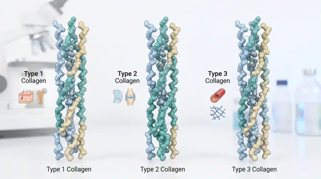
Collagen types 1, 2, and 3 make up about 80–90% of all collagen in the body. Type 1 supports skin and bone; type 2 supports cartilage and joints; type 3 supports blood vessels and internal organs. Knowing the difference between collagen type 1, 2, and 3—especially collagen type 1 vs 2—is essential for choosing the right type for any application.
What Are Collagen Types 1, 2, and 3?
Collagen types 1, 2, and 3 all belong to the fibrillar collagen family. This means they form fibers that give tissues their strength and structure. But each type has a different chain makeup and shows up in different parts of the body.
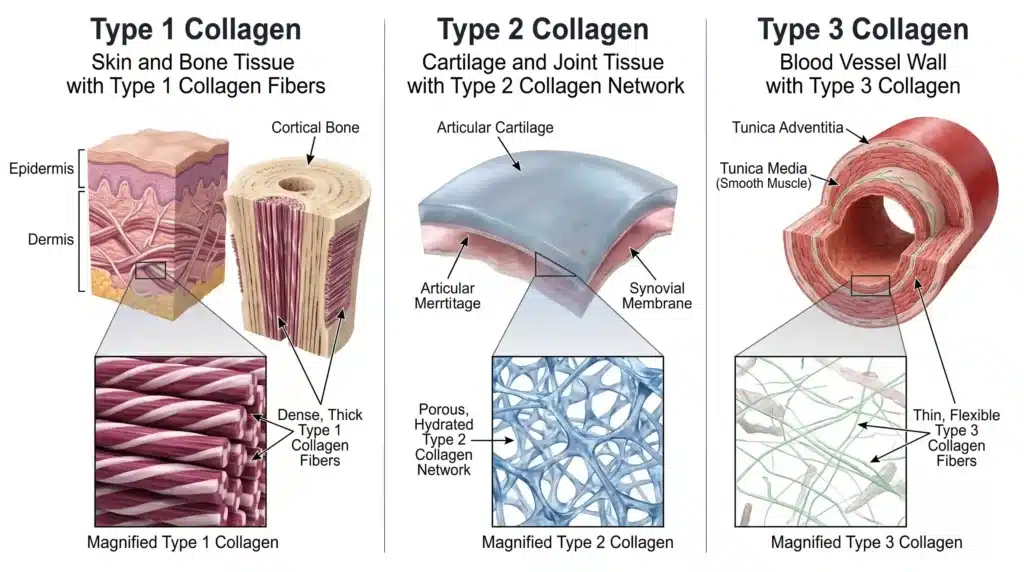
Type 1 collagen is made of two identical α1(I) chains and one slightly different α2(I) chain. It is the most common collagen in the body and is found in skin, bone, tendons, ligaments, and teeth. Its main job is to provide tensile strength and structural support.
Type 2 collagen is made of three identical α1(II) chains. It is mostly found in cartilage, making up 90 to 95 percent of the protein in joint cartilage. It also shows up in the eye and in spinal discs. Its role is to absorb shock and resist compression.
Type 3 collagen is also made of three identical chains, but of the α1(III) type. It often works alongside type 1 in skin, blood vessels, and organs. During wound healing, type 3 levels go up early on and are later replaced by type 1. It helps support hollow organs and blood vessel walls.
Together, these three types form the main structural framework of the body. But when people ask about collagen type 1 vs 2, they usually want to know which one supports skin and bone versus joints. Looking at collagen type 1 vs 2 vs 3 side by side helps make these differences clear.
How Their Chains Are Put Together
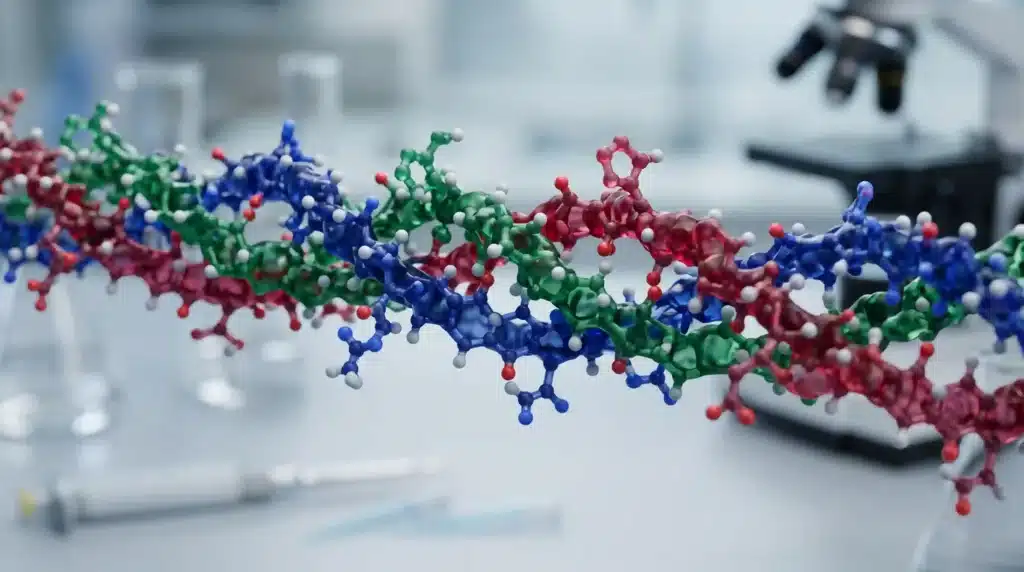
The basic difference between collagen type 1, 2, and 3 comes down to how their chains are arranged. This is where the collagen type 1 vs 2 comparison starts.
Type 1 collagen has two different kinds of chains — two α1(I) chains and one α2(I) chain. This mixed setup is unusual among fibrillar collagens. Its molecular weight is about 359 kDa. When you look at collagen type 1 vs 2 vs 3, type 1 stands out because it is the only one with two different chain types.
Type 2 collagen has three identical α1(II) chains. Type 3 shares this simpler structure, with three identical α1(III) chains. The structural difference between collagen type 1 and 2 is important: type 1 is the only major fibrillar collagen that uses two different chains.
All three types fold into a triple helix. Each chain has repeating Gly-X-Y patterns, where X and Y are usually proline and hydroxyproline. Glycine sits in the middle of the helix and makes tight winding possible. When these molecules form larger fibers, type 2 molecules are spaced further apart (16–17 Å) than type 1 (14 Å), mainly because type 2 has more sugar groups attached. Type 3 fibers are generally thinner and have fewer cross-links than type 1. These packing differences affect how stiff or flexible each type is: type 1 is tightly packed and rigid; type 2 is looser and more elastic; type 3 falls somewhere in the middle.
What Makes Their Amino Acids Different
The amino acid profiles of collagen types 1, 2, and 3 are fairly similar — glycine, proline, and hydroxyproline make up most of the content. But there are small differences in certain sequences and in how the chains are modified after they are made.
The Gly-Pro-Pro sequence helps stabilize the helix. Type 2 collagen has more of these stabilizing sequences than type 1, which is why type 2 can handle higher temperatures. Type 3 falls in between. This is one of the key differences between collagen type 1 and 2 that also affects how they are processed.
Lysine hydroxylation also varies across the three types. Type 2 gets more hydroxylation and glycosylation than type 1, which helps it hold more water. Type 3 has hydroxylysine levels similar to type 2 but with different glycosylation patterns. The ratio of hydroxyproline to hydroxylysine can be used in the lab to tell the three types apart.
When it comes to sugar content, type 1 has less than 0.5 percent carbohydrate by weight. Type 2 has much more because of its higher glycosylation. Type 3 is somewhere in the middle. These differences affect how much water each type holds, how its fibers form, and how it behaves in tissues.
Which One Handles Heat Better
Collagen types 1, 2, and 3 handle heat differently. Because type 2 has more Gly-Pro-Pro stabilizing sequences, it has a higher melting point than type 1. Type 1 melts at about 36.3°C, which is close to body temperature. Type 2 melts at a noticeably higher temperature. Type 3 behaves more like type 1, though the exact temperature can vary depending on the source.
This matters a lot in manufacturing. When making collagen peptides, temperature control is crucial. If the temperature is off, one type might break down while the other stays intact. For native type 2, even shipping in hot weather can cause problems if temperatures go above 40°C. Knowing the difference between collagen type 1 and 2 in terms of heat stability helps guide production and storage decisions.
Where They Are Found and What They Do
The most useful way to tell collagen types 1, 2, and 3 apart is by where they are located in the body and what they do there.
Type 1 collagen is the main structural protein. It supports the skin, makes bones hard, and helps tendons transmit force. It forms thick, strong fibers that resist pulling. About 90 percent of the collagen in the body is type 1. In the skin, type 1 keeps the dermis firm. As we age, production goes down and wrinkles start to form. In bone, type 1 provides the framework for minerals to deposit on. Without it, bones can’t harden properly.
Type 2 collagen is found almost entirely in cartilage. It forms thin, loosely woven fibers that absorb shock and handle compression. It is made by chondrocytes and makes up 95 percent of the protein in joint cartilage. Type 2 supports joints by providing a cushion that springs back after pressure is released. The genes for type 2 are only active in cartilage, the eye, and the spine.
Type 3 collagen is found alongside type 1 in skin, blood vessels, and organs. It is especially common in hollow organs like the uterus, liver, and arteries. Type 3 supports blood vessel walls and organs, and it plays a key role in wound healing — its levels go up early in the repair process and are later replaced by type 1. In the collagen type 1 vs 2 vs 3 comparison, type 3 is often overlooked, but it is vital for vascular and organ health.
These tissue-specific roles are what really set collagen type 1 vs 2 vs 3 apart.
Fiber Structure and Water Retention
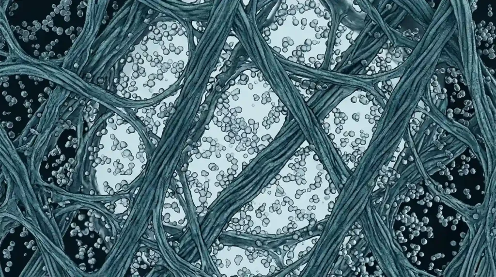
The structure of the fibers and how much water they hold also differ across the three types. Type 2 fibers hold 50 to 100 percent more water than type 1 fibers. This water is essential for shock absorption — when you walk or run, the water in cartilage helps spread out the impact. Type 3 fibers have water-holding properties closer to type 1, though with some variation.
Type 2’s ability to hold more water comes from its higher glycosylation and wider molecular spacing. Each glycosylation site can bind several layers of water, and the sugar chains form networks that make type 2 act like a sponge. Fiber diameter also varies: type 2 fibers are thinner and more flexible than type 1; type 3 fibers are also thin but have different cross-linking patterns. Type 2 has more cross-linking sites, which helps keep its network stable in cartilage.
Type 2 fibers join with glycosaminoglycans like chondroitin sulfate and hyaluronic acid to form large aggregates that trap water and create the load-bearing system of cartilage. This complex structure is unique to type 2 among collagen types 1, 2, and 3.
How Cells Respond to Each Type
Collagen types 1, 2, and 3 don’t just sit there as structural material — they also send signals to cells. When chondrocytes are grown on type 1 collagen, they flatten out and lose their cartilage-specific features. On type 2 collagen, they stay round and keep producing glycosaminoglycans. Cell growth also differs: on type 1 scaffolds, cells multiply early on but then drop off; on type 2, they stay steady. Type 3 has been found to promote blood vessel formation and support fibroblast activity in wound healing.
These differences matter in tissue engineering and regenerative medicine. Type 2 scaffolds are better at keeping chondrocytes functional, while type 1 is preferred for bone and skin applications. Type 3 is often used in vascular grafts and wound dressings. Some approaches combine types to get the best of each.
How They Are Used in Practice
In the real world, choosing between collagen types 1, 2, and 3 comes down to what tissue you are targeting.
Type 1 collagen is used for skin, bone, and tendon support. Most collagen supplements on the market are type 1, usually sourced from cow hide or fish scales. Hydrolysis breaks it down into small peptides that are absorbed and used by the body. These peptides, especially glycine-proline-hydroxyproline fragments, are known to stimulate skin cells.
Type 2 collagen is used for joint health. Native type 2 works through the immune system: it is recognized by immune cells in the gut, which then send signals to joints to reduce inflammation and protect cartilage. This is why the effective dose is so low — only 40 to 100 mg per day — compared to 5 to 10 grams per day for type 1. If type 2 is heated, it loses this special effect.
Type 3 collagen is used for vascular health and wound healing. It is often combined with type 1 to better mimic the natural matrix of skin and blood vessels. Some products include all three types to support skin, joints, and blood vessels at the same time.
The difference between collagen type 1 and 2 in dosage and how they work is especially important when formulating products.
Where They Come From and How They Are Made
Collagen types 1, 2, and 3 come from different raw materials and need different processing methods.
Type 1 is extracted from cow hide, cow bone, or fish skin and scales. Enzymes break it down into peptides of 2,000 to 5,000 daltons. Temperature must be controlled carefully to avoid turning it into gelatin.
Type 2 is almost always sourced from chicken sternal cartilage. The cartilage is concentrated there and easy to purify. After removing calcium and other non-collagen parts, controlled hydrolysis produces peptides that dissolve faster than native collagen. Chicken-derived type 2 often keeps its natural glycosaminoglycans, including chondroitin sulfate and hyaluronic acid, which adds nutritional value. At Atnutra, we focus on type 2 collagen peptides from high-quality avian sternum cartilage. Our controlled hydrolysis process breaks down the native structure into highly soluble peptides that dissolve quickly in water, supporting fast absorption. The product naturally contains a 3-in-1 matrix of type 2 collagen, chondroitin sulfate (≥25%), and hyaluronic acid (≥5%), giving formulators a complete cartilage-based ingredient without needing to source each component separately.
Type 3 is usually sourced from cows or pigs, often alongside type 1 because they coexist in skin and blood vessels. Pure type 3 is less common and more expensive.
The choice between native and hydrolyzed type 2 depends on the intended use. Native is more fragile and costly, while hydrolyzed is more stable and easier to work with.
Stability and Storage Considerations
Hydrolyzed type 1 peptides are very stable at room temperature as long as they are kept dry. Type 3 hydrolysates are similar. Native type 2 is more delicate — heat or too much mechanical force will unwind its triple helix and turn it into ordinary gelatin. That is why native type 2 often needs refrigerated storage and temperature-controlled shipping. Hydrolyzed type 2, like type 1, is stable under normal conditions.
This stability difference is an important practical factor when comparing collagen type 1 vs 2 for bulk sourcing.
Comparison Table
| Feature | Type 1 Collagen | Type 2 Collagen | Type 3 Collagen |
|---|---|---|---|
| Chain composition | α1(I)₂α2(I) (heterotrimer) | α1(II)₃ (homotrimer) | α1(III)₃ (homotrimer) |
| Primary location | Skin, bone, tendon, ligament | Cartilage, vitreous humour | Skin, blood vessels, organs |
| Primary function | Tensile strength, structural support | Shock absorption, compression | Organ support, vascular integrity |
| Proportion in body | ~90% of total collagen | >50% of cartilage protein | ~5–10% of total collagen |
| Fiber thickness | Thick | Thin | Thin |
| Water content | Lower | 50–100% higher | Intermediate |
| Glycosylation level | Low | High | Intermediate |
| Thermal stability | Melts ~36°C | Higher melting point | Similar to type 1 |
| Common source | Bovine hide, fish scales | Chicken sternal cartilage | Bovine/porcine skin |
| Typical daily dose | 5–10 g (hydrolyzed) | 40–100 mg (native) | Varies (often in blends) |
| Mechanism | Provides building blocks | Immune modulation | Structural support |
Additional Technical Details
Hydroxylation of proline and lysine happens after the protein chain is made. This process requires specific enzymes and vitamin C. Without enough vitamin C, the helix can’t form properly, leading to weak tissues — that is the basis of scurvy.
Type 1 has more hydroxyproline overall, but type 2 has more hydroxylysine and glycosylated hydroxylysine. This difference can be measured in the lab and used to confirm which type of collagen is present in a sample.
Cross-linking also differs between the types. Type 1 forms strong cross-links that make tendons tough. Type 2 forms more flexible cross-links that allow cartilage to be elastic. Type 3 has cross-links that support organ stretchiness. As we age, cross-links build up, making tissues stiffer.
After hydrolysis, each type produces its own pattern of peptide fragments. These can be used as fingerprints to check for purity and authenticity.
Summary
Collagen types 1, 2, and 3 are all fibrous and helical, but they differ in many ways — from how their chains are arranged to how they behave in the body. Type 1 is the stiff, structural type for skin and bone. Type 2 is the water-rich, elastic type for joints. Type 3 supports blood vessels and organs.
Understanding the difference between collagen type 1 and 2 — and where type 3 fits in — is key to picking the right ingredient. Whether you are looking at collagen type 1 vs 2 or considering all three, knowing these differences leads to better decisions.